An Accessible Approach to Optogenetics in the Classroom Using C. elegans
By Heather J. Rhodes
After the success I had using optogenetics with Drosophila in my undergraduate neurophysiology class last year (read, Implementing Optogenetics in the Classroom Part One: Drosophila), I decided to try working with another species — C. elegans — to conduct an optogenetics activity in my neuroscience senior seminar class this year.
Getting Familiar With a New Species
Like Drosophila larvae, C. elegans are transparent, which makes in vivo optogenetics very easy. By shining light of the wavelength that stimulates channel rhodopsin onto their bodies, you can observe behavioral changes.
Also, like Drosophila, you can order lines of C. elegans worms that express channel rhodopsin or other effectors in specific populations of neurons.
For my course, I used strain ZX460, which expresses channel rhodopsin in cholinergic neurons. Activation of channel rhodopsin in this line induces shortening of body length and a distinct coiling behavior.
Ordering and Maintaining C. elegans
Even as a C. elegans novice, I found this species was even easier to raise and maintain than Drosophila.
There is a huge repository of information on ordering and maintaining worms available online, particularly WormBook and the Caenorhabditis Genetics Center. To maintain worms, you need special agar plates seeded with a particular strain of E. coli. These agar plates can then be stored in an incubator or at room temperature.
Because I only needed to maintain a small number of plates for a couple of weeks, I contacted a C. elegans lab at a neighboring university. They provided me with a couple dozen plates and a small culture of E. coli.
All-trans retinol (ATR, Sigma R2500) is required for functional channel rhodopsin expression. I added ATR to the E. coli culture on some plates, leaving others without ATR to serve as a control group (as described in Goodman et al., 2014).
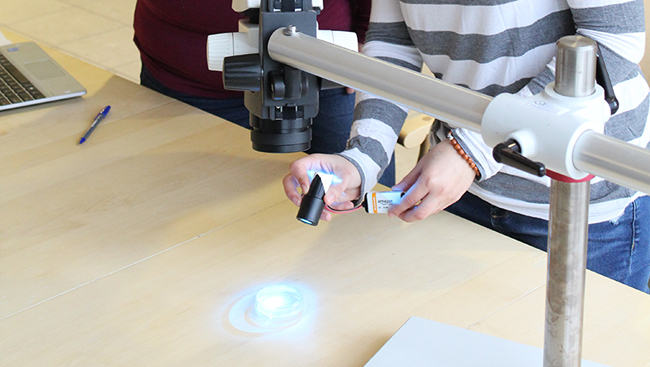
To deliver the appropriate light stimulus to activate the channel rhodopsin, I used a simple and inexpensive tool described by Titlow et al., 2014. It consists of a blue LED powered by a 9V battery, taped to a standard microscope eyepiece to focus the light.
Conducting the Experiment
Students then observed the C. elegans under a standard student dissecting scope.
I also supplied students with an inexpensive adapter that aligns a cell phone camera to the microscope eyepiece, allowing them to easily take photos and video of worm behavior.
To watch a demonstration of this experiment in progress, including requirement equipment and video footage of results, watch, Using Optogenetics with C. elegans for Inquiry-Based Student Experiments.
Leading an Inquiry-Based Student Activity
I developed an inquiry-based student activity (download the C. elegans lab handout) that could be shortened or expanded to fill a 50 minute class or a three hour lab. The activity was successful because it enabled students to observe changes in behavior driven by the optogenetic stimulation.
Students compared responses to control worms that were genetically identical but lacked the co-factor ATR. They were therefore able to see the contractile and coiling behaviors were driven by optogenetic activation of cholinergic neurons, independent of any photophobic behavior the light exposure may induce.
Students were also prompted to think about the function of these cholinergic neurons in C. elegans.
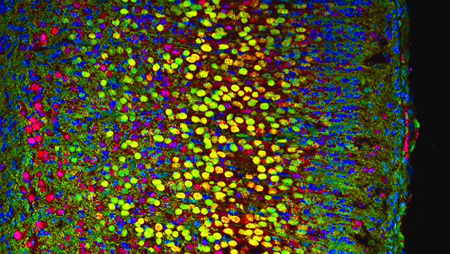
An additional component that could easily be added to this activity would be an examination of the anatomy of the cholinergic system within the worm. The cholinergic neurons in this line also express eYFP. If an appropriate fluorescent microscope is available, students could examine the location of labeled neurons, further expanding the scope of the laboratory exercise.
Supplies and Budget
- C. elegans strain ZX460: $7 (plus an annual $25 fee to use the CGC)
- NGM plates, E. coli: costs vary depending on quantities and needs
- ATR: approximately $50 for 25 mg (plus another $50 for shipping on dry ice)
- Ethanol (one ml) to dissolve ATR
- LED, battery, and snap connector: less than $10
- Soldering iron to connect LED to snap connector
- Microscope eyepiece with 10x lens: borrow from existing scope
- Dissecting microscope
- Microscope/smart phone adapter (optional): approximately $25
Download the C. elegans lab handout.
Visit the Community forum for all eight modules to share your insights and best practices, ask questions, and engage with other training series’ participants.
Complete this short survey to provide valuable feedback about Module 5 to SfN and the series faculty.


